SERVICES WE OFFER

Take the first step and find best treatment with us


WHAT IS CATARACT?

Cataract is a clouding of the eyes natural crystalline lens and is the most common cause of vision loss in older adults. Normally, light passes through the clear lens and is focused onto the retina at the back of the eye. With age, the lens gradually becomes more and more opaque and obstructs the passage of light, leading to a decrease in vision which affects day-to-day living and quality of life. Most patients notice symptoms of cataract after the age of 60, in one or both eyes. Some people, however, develop cataract in their 50s and even their 40s.

Most cataracts develop slowly and your eyesight may not get affected in the initial stages. However, with time, the clouding of your lens will eventually interfere with your vision. When the cataract is the size of a pin head, the cloudiness affects only a small part of the lens. You may notice a slight change in your vision. As it grows larger, the cataract clouds more of your lens. This scatters and blocks the light as it passes through the lens, preventing a sharply defined image from reaching your retina. This may lead to more noticeable symptoms.

Most cataracts develop slowly and your eyesight may not get affected in the initial stages. However, with time, the clouding of your lens will eventually interfere with your vision. When the cataract is the size of a pin head, the cloudiness affects only a small part of the lens. You may notice a slight change in your vision. As it grows larger, the cataract clouds more of your lens. This scatters and blocks the light as it passes through the lens, preventing a sharply defined image from reaching your retina. This may lead to more noticeable symptoms.


RISK FACTORS OF CATARACT

Other than aging, factors that increase your risk of cataracts include a previous eye injury or inflammation, a previous eye surgery, excessive exposure to sunlight (ultraviolet-B rays), medical conditions such as diabetes and high blood pressure, genetic abnormalities, skin diseases, smoking, ionizing radiation (treatment for cancer) and prolonged use of corticosteroid medications.

RATE OF GROWTH

It is not always possible to predict exactly how fast a cataract will develop as the rate of growth varies from person to person and may also vary between both eyes in a particular individual. There are many kinds of cataracts, and they can either develop very slowly or progress rapidly. Cataracts that are caused by the aging process most often progress gradually over a period of years. Cataracts that affect diabetics and young people, or cataracts that are caused due to certain injuries or use of certain medications may progress rapidly over a few months.

    CLASSIFICATION OF ADULT CATARACTS:

  • An adult cataract can be immature, mature, or hyper-mature.
  • When there are some remaining clear areas in the lens, the cataract is considered as immature.When the lens is completely opaque, the cataract is onsidered as mature.
  • When the cataract has a leaky fluid, it is considered as hypermature.

TYPES OF CATARACT

The different types of cataract include:

Nuclear cataract -This type of cataract forms in the center of the lens. Initially this cataract may cause a temporary improvement in your reading vision or more near-sightedness. With time, however, it turns the center of the lens yellow or even brown, leading to clouding of vision and difficulty distinguishing between shades of colour.

Cortical cataract -This type of cataract forms at the edges of the lens. It begins as whitish streaks or wedge-shaped cloudiness around the outer edge of the lens cortex and slowly extends to the center of the lens with time. This interferes with light passing through the center of the lens. People with diabetes are more prone to develop this type of cataract.

Posterior subcapsular cataract -This type of cataract affects the back of the lens and progresses much faster than other types of cataract. This cataract starts as a small, opaque area that generally forms near the back of the lens, right in the path of light. It interferes with your reading vision, reduces your vision in bright light, and causes glare or halos around lights at night. People with diabetes , Uveitis long term steroid either in the form of drops or tablets, retinitis pigmentosa may have a higher risk of developing this type of cataract.

Congenital cataract -This type of cataract is present at birth or develops during childhood, often in both eyes. Congenital cataracts may be genetic, or may occur due to infection, inflammation, trauma, metabolic problems or drug reactions. These cataracts may be extremely small and don’t always affect vision. However, moderate to severe cataracts that affect vision may need to be treated with cataract removal surgery at the earliest to avoid lazy eye (Ambylopia).

Traumatic cataract -This type of cataract may develop following injury to the eye while playing sports such as tennis, cricket, badminton, during an accident or due to the use of blunt force, which results in damage to the lens inside the eye. This kind of cataract may vary from minor type of opacification of the natural lens, to a profound damage to the natural lens, along with injury to outer layers of the eye and the posterior part of the eye as well.

Secondary cataract -This type of cataract is also known as posterior capsular opacification (PCO) or after-cataract. Some months or years after cataract surgery, a small percentage of people experience a decline in their eyesight. The posterior portion of the lens capsule (the part behind the natural lens) is left inside the eye during cataract surgery, and the IOL is implanted directly in front of it.This cataract makes the back surface of the lens opaque or cloudy, causing vision to become blurred again.


  • Cloudy or blurred vision,
  • Distorted or double vision.
  • Sensitivity to light and glare.
  • Difficulty seeing in poor lighting conditions.
  • SICS-Small Incision cataract surgery
  • Phaco/Phacoemulsification
  • Micro Incision (1.8 mm, MIOL)
  • Monofocal Intraocular Lens
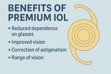
  • Provides enhanced vision for near and far .
  • It makes you spectacle free most of the time.
  • This lens is helpful if you are constantly switching distances during the day and greatly reduces the need for wearing glasses or contact lenses after surgery doesn't give good intermediate vision.
  • Provides distance vision for watching TV and other far activities.
  • Provides intermediate vision for working on computers/laptops.
  • Provides near vision for reading, writing, seeing mobile.
  • Phacoemulsification refers to modern cataract surgery via a small 2-3 mm cut, in which a handheld probe is used to break up and emulsify the lens into liquid using ultrasound energy. The resulting emulsion is sucked out. After these steps, the artificial foldable intraocular lens is inserted inside the eye.
  • Requires a small incision of 3.0 mm
  • Stitch less surgery
  • Cataract is emulsified and removed using an ultrasonic phacoemulsification probe
  • Requires a very small incision of less than 2mm
  • Cataract is emulsified into small pieces by phacoemulsification and a foldable MIOL is implanted
  • Walk-in, Walk-out procedure
  • Stitch less, bloodless, painless surgery

WHICH INTRAOCULAR LENS IS BEST?

You cataract surgeon can help you decide which intraocular lens (IOL) is best for you based on your daily activities and which type of vision, near or distance focus, is required to complete those activities. Other factors to consider include pre-existing conditions such as astigmatism, glaucoma, macular degeneration, corneal disease, or other conditions which will need a specific type of lens.

POTENTIAL COMPLICATIONS OF CATARACT SURGERY

While any kind of surgery entails a certain amount of risk, cataract surgery is highly successful and generally considered as one of the safest surgeries you can have. Most complications are minor, such as swelling of the cornea or retina, increased pressure in the eye, and droopy eyelid. In general, the risk of severe visual loss is very rare, but may occur as a result of infection or bleeding inside the eye, or even retinal detachment, which may occur months or years after a perfectly successful cataract surgery. Underlying eye damage from other eye diseases such as glaucoma, diabetic retinopathy, or macular degeneration or pre existing co morbid conditions of the eye can increase your risk of complications and reduce the chance of improved vision after surgery.

Refractive Errors

Normal Eye Light rays enter the eye through the cornea where they are refracted (bent) and pass through the pupil to finally form a sharp focus or image on the innermost sensitive layer of the eye, the retina, just like in the camera. The retina then sends this information to the brain via the optic nerve and the brain perceives the final image. When the image is exactly formed on the retina and there is no power in the eyes, it is called Emmetropia.

Refractive errors are eye disorders in which the light is not properly refracted to a point focus on the retina and instead a blurred image is perceived.

Refractive errors

Types of Refractive Errors

Refractive errors are eye disorders and not diseases. These include myopia, hyperopia and astigmatism. Most people have one or more of them.

How to diagnose?

The common method of measuring vision loss is the vision chart. Refraction and retinoscopy can help detect the actual error and the spectacle power.

Refractive errors

Myopia (Nearsightedness)

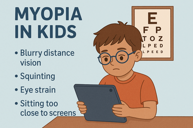
Nearsightedness or myopia is a condition in which near objects are seen clearly, but distant objects are not clear. This occurs due to light rays focus in front of the retina due to either longer eye ball or increased corneal curvature of a steep cornea. Myopia occurs in different degrees from minimal to extreme. The more myopic you are the blurrier your vision is at a distance and objects will have to be closer to you so you can see them clearly.

Nearsightedness is a common vision condition affecting nearly 20% of the Indian population. Commonly it starts in school-age children. Because the eye continues to grow during childhood, nearsightedness may increase up to the age of 18 to 21, which generally stabilizes by then. Recent studies showed that the nearsightedness could be hereditary as well as could be caused by too much stress on the eyes in terms of very close vision work in growing children. However, larger studies are still going-on to prove these theories.

Refractive errors

A sign of nearsightedness is difficulty in seeing distant objects like TV screen and the child may want to watch it from very near or difficulty in seeing blackboard in the school, for which the child may want to copy from the student sitting next instead of looking at the blackboard. These children may have poor class work or class notes in spite of being regular and good at home work. A comprehensive eye examination is required in such cases to rule out nearsightedness.

It can be corrected by prescribing eyeglasses or contact lenses to optically correct nearsightedness, which you may only need to wear for certain activities, like watching TV or a movie or driving a car, or they may need to be worn continuously.

One thing that needs to be remembered is that in case of high myopia, there is a risk of retina getting detached, which would require a surgical treatment. Hence, a regular follow-up with eye doctor is a must.

Hyperopia (Farsightedness)

Farsightedness, or hyperopia is a condition in which distant objects are usually seen clearly, but close ones are not clear. Farsightedness occurs if your eyeball is shorter than the normal or the cornea is less curved than normal or flat, so light entering your eye is focused behind the retina. It is usually inherited. A child is usually born with hyperopia and as the eye grows it reduces.

Hyperopia

Common signs of farsightedness include difficulty in clearly seeing near objects, headaches, eye strain, and/or fatigue after close work. Although the hyperopia is not as common as myopia the common vision screenings, often done in schools, are generally ineffective in detecting this condition. A comprehensive ophthalmological examination is required in all those with above mention complaints. In mild cases of hyperopia, patient may not need corrective glasses, as eyes may be able to compensate by working harder. In other cases, your ophthalmologist may prescribe spectacle or contact lenses to optically correct this condition.

Astigmatism (Distorted Vision)

Astigmatism is a condition where the front surface of your eye, the cornea, is irregular in shape that is not perfectly round but more oval preventing the light to focus at one point on the back of your eye, the retina. As a result, the vision would be blurred at all distances. Astigmatism rarely occurs alone. It is usually accompanies myopia or hyperopia.

Astigmatism

Most of astigmatism can be corrected with properly prescribed and fitted eyeglasses and/or contact lenses. However, higher astigmatism may be better handled by surgical means like astigmatic keratotomy or LASIK.


Types of Refractive Surgeries

Removing the natural lens and replacing it with an IOL of adequate power. It is similar to a routine phacoemulsification surgery, except that it is done in a clear lens and not a lens with cataract.

Keratoconus is a condition where cornea becomes ecstatic & vision of patients deteriorates. We have state of art facility for collagen cross linking using imported Riboflavins which changes intrinsic properties of cornea.

This is performed to correct remaining spectacle power in patients where IOL is already done somewhere else

In this We put a Multifocal IOL or Toric Multifocal IOL after removing Crystalline lens for Presbiopia Treatment.


Retina & Vitreous

Retina is the light sensitive inner layer of the eye where the images are formed and are later relayed to the brain. This is very important for vision. For investigation of retina and vitreous diseases we have Fundus Camera (Carl Ziess, Germany), OCT (Carl Ziess, Germany). We also have facility of Green Laser (Carl Ziess, Germany) for doing PRP and Endo-Laser. We also have facilities for various Intra Vitreal Injections like Lucentis, Avastin, Ozudex and Vancomycin etc.

Diabetic Retinopathy

Diabetic retinopathy, the most common diabetic eye disease, occurs when blood vessels in the retina change. Sometimes these vessels swell and leak fluid or even close off completely. In other cases, abnormal new blood vessels grow on the surface of the retina
Diabetic retinopathy usually affects both eyes. People who have diabetic retinopathy often don�t notice changes in their vision in the diseases early stages. But as it progresses, diabetic retinopathy usually causes vision loss that in many cases cannot be reversed.

Diabetic eye problems

There are two types of diabetic retinopathy:
Background or nonproliferative diabetic retinopathy (NPDR)

Nonproliferative diabetic retinopathy (NPDR) is the earliest stage of diabetic retinopathy. With this condition, damaged blood vessels in the retina begin to leak extra fluid and small amounts of blood into the eye. Sometimes, deposits of cholesterol or other fats from the blood may leak into the retina.

    NPDR can cause changes in the eye, including:

  • Microaneurysms: small bulges in blood vessels of the retina that often leak fluid.
  • Retinal hemorrhages: tiny spots of blood that leak into the retina.
  • Hard exudates: deposits of cholesterol or other fats from the blood that have leaked into the retina.
  • Macular edema: swelling or thickening of the macula caused by fluid leaking from the retina�s blood vessels. The macula doesn�t function properly when it is swollen. Macular edema is the most common cause of vision loss in diabetes.
  • Macular ischemia: small blood vessels (capillaries) close. Your vision blurs because the macula no longer receives enough blood to work properly.

Proliferative diabetic retinopathy (PDR)

Proliferative diabetic retinopathy (PDR) mainly occurs when many of the blood vessels in the retina close, preventing enough blood flow. In an attempt to supply blood to the area where the original vessels closed, the retina responds by growing new blood vessels. This is called neovascularization. However, these new blood vessels are abnormal and do not supply the retina with proper blood flow. The new vessels are also often accompanied by scar tissue that may cause the retina to wrinkle or detach.
PDR may cause more severe vision loss than NPDR because it can affect both central and peripheral vision. PDR affects vision in the following ways:

Delicate new blood vessels bleed into the vitreous � the gel in the center of the eye � preventing light rays from reaching the retina. If the vitreous hemorrhage is small, you may see a few new, dark floaters. A very large hemorrhage might block out all vision, allowing you to perceive only light and dark. Vitreous hemorrhage alone does not cause permanent vision loss. When the blood clears, your vision may return to its former level unless the macula has been damaged.

Scar tissue from neovascularization shrinks, causing the retina to wrinkle and pull from its normal position. Macular wrinkling can distort your vision. More severe vision loss can occur if the macula or large areas of the retina are detached.

If a number of retinal vessels are closed, neovascularization can occur in the iris (the colored part of the eye). In this condition, the new blood vessels may block the normal flow of fluid out of the eye. Pressure builds up in the eye, a particularly severe condition that causes damage to the optic nerve.

Vitrectomy

Vitrectomy is the surgical removal of the vitreous gel from eye. It may be done when there is a retinal detachment, because it gives your eye doctor (ophthalmologist) better access to the retina of the eye. The vitreous gel may also be removed if blood in the vitreous gel (vitreous hemorrhage) does not clear on its own.

Buckling Surgery for Retinal Detachment

Scleral buckling surgery is a common way to treat retinal detachment. It is a method of closing breaks and flattening the retina. Scleral buckling is effective in supporting a tear, hole, or break in the retina that has caused the detachment. It is rarely helpful on its own when scar tissue tugging on the retina has caused the detachment (traction detachment).

Glaucoma Treatment

Glaucoma is a serious eye disease caused by increased fluid pressure within the eye, which can damage the optic nerve that transmits images to the brain. One of the leading causes of irreversible blindness, the estimated number of cases of glaucoma in India is 12 million. This is around one fifth of global burden of glaucoma.
Glaucoma is known as the “silent thief of sight” as it is a symptomless disease, and people usually do not realize that they are suffering from glaucoma unless they visit the ophthalmologist or have reached an advanced stage of disease. If undiagnosed and untreated, glaucoma can lead to irreversible loss of vision and even cause blindness. Though the disease is not curable, the progress of glaucoma can be halted and total blindness can be prevented if it is detected early.


Glaucoma
Glaucoma

HOW GLAUCOMA AFFECTS VISION

In glaucoma the fluid pressure inside the eye increases, and this leads to progressive damage of the optic nerve, resulting in a gradual loss of vision. Initially, the loss of vision starts from periphery and progressively affects the central vision. The central visual acuity is affected only in the advanced stage.
The front half of the eye is filled with a clear fluid known as the “aqueous humor”. This fluid is continuously produced and constantly flows in and out of the eye, maintaining a steady pressure inside the eye. If there is either reduced drainage or excessive production of this fluid, the pressure in the eye will increase. This increased eye pressure, if left untreated, can damage the eye (optic) nerve and cause permanent vision loss. Not everyone with high eye pressure will develop glaucoma, and some people with normal eye pressures may also develop glaucoma.
The disease usually affects both eyes, though one may be more severely affected than the other. Unfortunately, the damage to the eye nerve due to glaucoma is irreversible and the loss of vision cannot be reversed. However, this condition can be halted and the remaining vision can be retained if glaucoma is diagnosed on time and appropriate treatment is started.

TYPES OF GLAUCOMA

The different types of glaucoma include:

Primary open angle glaucoma – is the most common type of glaucoma. In this type of glaucoma, the part of the eye through which the fluid of the eye flows out is open, permitting the outflow of fluid, but the patient still has high pressure. This type of glaucoma develops slowly without any symptoms. Initially it affects the peripheral or side vision and very gradually progresses to the centre. This is the reason why many people are not aware that they have the condition until they have significant vision loss affecting central vision.
Angle closure glaucoma or closed angle glaucoma –is a less common type of glaucoma which occurs due to narrow drainage channels in the eye. Gradual closing of the angle is called chronic angle closure and if the drainage angle closes suddenly, it causes an acute angle closure attack. Acute angle closure glaucoma usually presents as an emergency. A patient who is in an acute angle closure attack will have symptoms of eye pain, nausea, vomiting, redness and blurred vision due to a rapid increase in the eye pressure. In such cases the patient needs immediate treatment by an eye specialist.
Normal tension glaucoma /Low tension glaucoma –In this type of glaucoma the optic nerve can get affected even though the pressure in the eye is normal. Although its cause is not entirely known or understood, normal tension glaucoma is believed to occur either because of an extremely fragile optic nerve that can get damaged even though the pressure in the eye is normal, or because of reduced blood flow to the optic nerve. Because of its silent nature, people usually do not have any visual complaints until a very advanced stage of the disease.
Secondary glaucoma –There are certain other types of glaucoma where there is an identifiable cause for increased eye pressure resulting in optic nerve damage and vision loss. These are called secondary glaucoma. It may be caused by prolonged, indiscriminate use of steroids, severe diabetic retinopathy, injuries to the eye, inflammation of the eye (uveitis) or advanced cases of cataract.
If you believe you have any of these risk factors get an eye examination done. Always remember to inform your eye doctor about the risk factors that you have. This will help your doctor decide how often you need to get your eyes examined.

GLAUCOMA IS HEREDITARY

Primary open-angle glaucoma, which is the most common type of glaucoma, is hereditary. If members of your immediate family such as a parent or sibling have glaucoma, you are four to nine times more likely to develop this disease than the rest of the general population.
Know your risks! Save your vision!
Be informed about your family’s medical history and understand the hereditary risks of glaucoma. Speak to your doctor if you have a family member with glaucoma, it may save your sight. Be vigilant about eye check-ups and alert to possible glaucoma symptoms.
Since early symptoms or pain may not show up in people with glaucoma, this disease is often diagnosed at a later stage when visual loss has already happened. If you’re over the age of 40 and have a family history of glaucoma, you should get a comprehensive eye exam, which includes dilated eye exams and eye pressure checks every year, even if your vision is healthy.
Without treatment, glaucoma can cause total permanent blindness within a few years. As this damage is irreversible, loss of vision due to glaucoma cannot be regained. However, if detected early, vision loss can be avoided, or delayed with the use of eye drops, laser treatment or surgery. Even a small reduction in pressure can make a big difference in slowing vision loss.

WHAT ARE THE TESTS I NEED TO UNDERGO?

The following tests help in finding out the presence of glaucoma and also its progression in the subsequent follow-ups.
Tonometry is a diagnostic test that measures the fluid pressure, known as intraocular pressure (IOP), inside your eye. The tonometry test is important as it can help your doctor evaluate whether or not you may be at risk of glaucoma.
Gonioscopy is a painless eye examination of the front portion of your eye (anterior chamber), to examine whether the area where fluid drains out of your eye (drainage angle) is open or closed. This test is important as it helps your doctor to diagnose and monitor various eye conditions associated with glaucoma.
The Visual Field test is a method of measuring your peripheral or side vision (which is affected first by glaucoma), through which your doctor can diagnose and monitor glaucoma. The data from the test is used to determine the severity of your glaucoma, level of vision loss, damage to the visual pathways of the brain, and other optic nerve diseases.
Ophthalmoscopy

GLAUCOMA PREVENTION

In today’s world, people are quite aware about how lifestyle choices affect our overall health, and the same goes for glaucoma. Lifestyle choices can, to some extent, influence eye pressure and affect the risk of developing glaucoma, but there aren’t enough proven studies about the same in glaucoma patients. However, a shift towards a healthier lifestyle makes for an overall positive change.
Exercise – Research indicates that regular exercise and an active lifestyle can help reduce their risk of glaucoma. Activities which raise your pulse by 20 to 25, like walking briskly, can help reduce IOP. The more number of steps a person walks throughout the day, less likelihood of an increase in IOP. Aerobics is known to cause a temporary decrease in the intraocular pressure but it has not been tested in glaucoma patients. Swimming reduces vulnerability of the optic nerve to an increase in the intraocular pressure.
Diet –Various studies have been undertaken to study the role of diet in glaucoma. A diet rich in green leafy vegetables is supposed to lower the eye pressure. Dietary nitrates in green leafy vegetables lowers risk of developing open angle glaucoma.
DHA (docosahexanoeic acid) is an omega- 3 fatty acid and it is helpful in preserving a healthy retina and lowering the IOP. Rich sources of DHA such as salmon, shellfish, tuna should be consumed twice or thrice a week. If its natural addition to the diet is not possible, DHA supplements in the form of fish oil supplements can be taken.
A diet rich in lutein and zeaxanthin helps in lowering intraocular pressure. It avoids oxidative damage to the optic nerve. Spinach, broccoli and sprouts are rich sources of lutein and zeaxanthin.
Antioxidants provide nutrients and strengthen the muscles and nerves in the eye. Rich sources of antioxidants are blueberries, grapeseed extract, goji berries, pecan nuts and cranberries.

What to avoid

Exercise – Lifting of heavy weights is known to cause a temporary increase in the intraocular pressure. Hence, patients who already suffer from glaucoma are advised to avoid lifting very heavy weights. Yoga, which is a very popular form of exercise includes various ‘asanas’ or positions. Some of these asanas, particularly those with the head – down position (sheershasana) have been proven to cause an increase in the eye pressure. Therefore, patients with glaucoma are advised to avoid such positions.
Diet –Foods which are rich in trans-fats, the kind found in deep fried food, prevent the optimal functioning of omega 3- fatty acids and increases eye pressure. Such food should be avoided.
Habit forming substances –such as caffeine, alcohol, tobacco and marijuana are known to have a negative effect on eye pressure. Caffeine, which is consumed widely, is known to cause a temporary increase in the eye pressure. Consumption of caffeine in small quantities is alright, but an excessive intake of caffeine is has been proven to increase the risk of glaucoma. Alcohol is known to decrease the eye pressure for a short duration but a daily consumption of alcohol is supposed to increase the risk of glaucoma. Smoking cigarettes is known to increase the risk of glaucoma. Though marijuana is known to lower eye pressure, it is not recommended for treatment due to its side effects.
Musical instruments –such as the trumpet and the saxophone are known to cause an increase in the eye pressure. Glaucoma patients are advised to avoid playing such high resistance wind instruments which can increase the intraocular pressure.

TIPS to make the life of a glaucoma patient easier

A better understanding of your treatment and medications can make it easier to live with glaucoma and easier for your doctor to control your disease. Early diagnosis, and therefore early intervention, is crucial in delaying the progression of the disease process.

Before the disease is detected:

Prevention is better than cure

  • Ophthalmic consultation is a must for everyone over the age of 40, in an eye hospital rather than getting tested for near vision at an optical shop.
  • Get glaucoma screening done if you have a family history of glaucoma, diabetes, hypertension, heart disease, asthma, arthritis, migraine, thyroid disease, using minus or plus powered glasses, any history of trauma to eye. If detected in the early stage, the disease can be arrested and existing vision can be retained.
  • Babies born with whitish coloured eyes or bigger than normal eyes, or babies with severe watering and difficulty in opening their eyes at birth, should be screened for congenital glaucoma.

After the disease is detected:

If you have undergone any surgery or laser treatment for glaucoma, it is important to understand that these procedures are performed to reduce the intra-ocular pressure and are an attempt to restore the remaining vision.

  • Follow the instructions of your doctor and attend follow up examinations as advised
  • Follow the technique properly while instilling the medication into the eye so that the desired effect of medication is attained.
  • Regular usage of medications
  • keeping alarms on your mobile phones will remind you to instil your medications on time
  • making a simple drug chart in a pocket diary as shown in the figure so that it can be noted and showed to your doctor as well.
  • If you have any difficulty in instilling the medication or if you have any discomfort with the medication, let your doctor know about it rather than stopping the medication.
  • Never stop the medications without consulting your doctor – people usually think that there is no change in vision and stop the medication. This can compromise your vision further.

The Pediatric Ophthalmology service of Saraswati Eye Care Centre has started with a noble aim of bringing back a smile to a child’s face. Over the years the department has grown to be a major referral centre for pediatric eye disorders. At present, this service involves not only diagnosis and management of various facets of pediatric eye disorders like refractive error, childhood cataract, glaucoma, amblyopia, squint, retinal disorders, congenital anomalies etc.. but also adult patients with squint/ double vision problems. The department provides appropriate therapeutic intervention for all stages of retinopathy of prematurity, a blinding disease of the premature newborn.


Surgeries

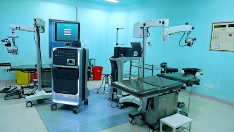
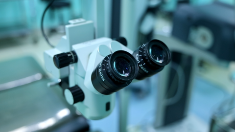
Saraswati Eye Care Centre provides a very child friendly ambience. We have a dedicated, state of art operating room for pediatric eye surgery with microscopes, vitrectomy machines etc.

    Various Surgeries like:

  • Correction of vertical and horizontal squints.
  • Glaucoma surgeries like trabeculotomy, trabeculectomy, drainage implants.
  • Simple and complicated pediatric retinal surgeries.
  • Cataract (cataract surgery with or without primary posterior capsulotomy and/ or IOL implantation).
  • Childhood eye injuries.
  • Minor procedures like foreign body removal and chalazion etc

Using the expertise of excellent anesthesiologists at Saraswati Eye Care Centre, we are equipped to perform ocular surgeries with ease in newborns especially those with multisystem ailments and at a very early age.
In short, all the possible diagnostic and treatment facilities are made available to the children of all strata under a single roof.

Corneal Blindness

Corneal blindness is the fourth leading cause of blindness in India. A majority of such patients are children. Treating corneal blindness is possible through cornea transplant. In this treatment, a healthy cornea is transplanted in place of a diseased cornea in entirety through (penetrating keratoplasty) or in part (lamellar keratoplasty). Since artificial corneas are not available, corneas have to be harvested only through eye donation.
The cornea is the eye�s outermost layer. It is the clear, dome-shaped surface that covers the front of the eye. It is responsible for more than 2/3rd of the eye�s focusing power. Unlike most tissues in the body, the cornea contains no blood vessels to nourish or protect it against infection. Instead, the cornea receives its nourishment from the tears and aqueous humor that fills the chamber behind it. To see well, all layers of the cornea must be free of any cloudy or opaque areas. When cornea becomes cloudy due to disease, injury, infection or malnutrition, vision is significantly lost or reduced.

The common corneal disorders are

Refractive errors

about-author If the cornea is flatter than normal or the eye is short, rays of light are focused behind the retina and causes hyperopia or farsightedness where close objects appear blurred. Astigmatism is a condition in which the uneven curvature of the cornea blurs and distorts both distant and near objects. The cornea is more curved in one direction than in the other. This causes the rays of light to focus on two separate areas of the retina, distorting the visual image. Refractive errors are usually corrected by eye glasses or contact lenses. Although these are safe and effective methods for treating refractive errors, refractive surgeries are becoming an increasingly popular option.

Allergies

about-author

Allergies affecting the eye are relatively common. They are most commonly related to pollen and dust in the air. They are usually immediate or delayed hypersensitivity reactions. Symptoms can include redness, itching, and burning, tearing, stinging and watery discharge. An increasing number of eye allergy cases are related to medications and contact lens wear.


Conjunctivitis (red / pink eye)

about-author

Conjunctiva is a translucent mucous membrane which lines the posterior surface of the eyelids and anterior aspect of the eyeball. A group of diseases that cause swelling, itching, burning and redness of the conjunctiva are termed as conjunctivitis. It is an inflammation of the conjunctiva associated with a discharge which may be watery, mucoid, mucopurulent or purulent. It can spread from one person to another if proper precautions are not taken.

Infections

A breach in the normal epithelial surface of the cornea associated with necrosis of surrounding corneal tissue is termed as corneal ulceration. Two main factors are responsible in the production of a corneal ulcer: damage to corneal epithelium and infection of the eroded area. They usually cause pain, redness, watering, discharge, photophobia i.e. intolerance to light and blurred vision. It can be caused by bacteria, fungi, viruses, acanthamoeba and many other organisms.

Dry Eye

Dry eye can be caused due to any of the following reasons:

  • Aqueous tear deficiency
  • Mucin deficiency
  • Lipid deficiency
  • Impaired eyelid function
  • Corneal epithelium abnormalities.

Corneal dystrophies affect vision in widely differing ways. Some cause severe visual impairment, while a few cause no vision problems and are discovered during a routine eye examination. Other dystrophies may cause repeated episodes of pain without leading to permanent loss of vision.

Keratoconus is characterized by progressive thinning and ectasia which results in deterioration of the quality of vision and also the quality of life. A new modality of treatment, based on collagen crosslinking with the help of Ultraviolet A (UVA, 365nm) and the photosensitizer riboflavin phosphate has been described which changes the intrinsic biomechanical properties of the cornea, increasing its strength by almost 300%.

Pterygia are more common in sunny climates and in the 20-40 age group. Scientists do not know what causes pterygia to develop. However, since people who have pterygia usually have spent a significant time outdoors, many doctors believe ultraviolet (UV) light from the sun may be a factor. In areas where sunlight is strong, wearing protective eyeglasses, sunglasses, and/or hats with brims are suggested. While some studies report a higher prevalence of pterygia in men than in women, this may reflect different rates of exposure to UV light.

Patients with ocular surface diseases suffer from loss of vision, discomfort, infection, erosions, ulceration, and destruction with scarring of the eye surface. The most common cause of these problems is the imbalance in the neural regulation which leads to an �unstable tear film�. Ocular surface failure manifests in two ways � the first one is the Limbal Stem Cell Deficiency in which the corneal epithelium is replaced by the conjunctival epithelium. In the second one, the corneal or the conjunctival epithelium changes with keratinisation and loss of mucosal epithelial characteristics.

A contact lens wearer who presents with conjunctival irritation and associated symptoms � such as itching, burning or tearing � may be suffering from any one of a number of conditions (such as allergy) of which the lenses may not be the primary cause. In other cases, however, contact lens wear itself is responsible for the condition.

Contact lens related problems

A contact lens wearer who presents with conjunctival irritation and associated symptoms � such as itching, burning or tearing � may be suffering from any one of a number of conditions (such as allergy) of which the lenses may not be the primary cause. In other cases, however, contact lens wear itself is responsible for the condition.


The department of Uveitis and ocular immunology armed with three qualified consultants, a well equipped clinical laboratory and state of the art ophthalmic diagnostic equipment deals with the whole battery of ocular inflammations including autoimmune diseases and infections in both adults and children.
The Uveitis & Ocular Immunology Service provides a multidisciplinary approach to the treatment of ocular inflammatory disease and other infectious disorders. Experts here deal with diagnosis and management of complex infectious and autoimmune diseases of the eye as well as eye diseases associated with various other systemic medical disorders. The diagnostics are backed by a well equipped laboratory, pathology and molecular diagnostic services, and the consultants are conversant with appropriate intervention techniques.
We co-ordinate with the other subspecialities of ophthalmology and internal medicine efficiently to get the optimal treatment outcomes for patients with complex Uveitic disorders.

What is Uveitis

Uvea is the middle part of the three coats of the eye. This further consists of the iris, ciliary body and the choroid. Inflammation of any of these parts is termed uveitis.

Types of uveitis

Based on the part of the uvea involved, uveitis may be Anterior (involving the iris), Intermediate (involving the ciliary body), Posterior (involving the choroid) or Panuveitis (involving all the parts).

What are the symptoms of uveitis?

Symptoms of uveitis are highly variable and may include any of the following: redness, pain, watering, inability to see bright light, floaters, and / or decreased vision.

What causes uveitis?

Uveitis occurs as a result of an immune reaction by our body to antigens (substances our body considers foreign). This reaction may occur against infectious agents such as bacteria, fungi, viruses and even parasites. In a small subset of patients, uveitis can occur due to undeterminable causes.

Investigations required for uveitic patients

Uveitic patients often require a whole battery of investigations in order that the underlying cause of uveitis be determined and hence appropriately treated. These usually include blood and urine tests and/ or X rays. At times, a sample of the fluid from the patients’ eye may have to be subjected to lab tests.

How is uveitis treated?

Steroids are the mainstay of treatment in uveitis. Depending on the location and the severity of the inflammation, they are used in the form of eye drops, eye ointment, injections around/ in the eye or injectable / oral medications. Anterior (and intermediate) uveitis is treated with topical steroids along with dilating eye drops which help in reducing the pain associated with inflammation. These drops are to be used until the inflammation has completely subsided. The dose, strength and duration of the drops are determined by your doctor who decides the treatment in accordance to the amount of inflammation.
Injection of the steroid around the eye (periocular steroids) is used in certain cases of intermediate uveitis (or in macular edema as a consequence of uveitis). This results in slow release of the drug over a period of three to four weeks.
Injectable/oral steroids are often indicated in posterior/panuveitis.
Besides steroids, the other group of drugs used in the treatment of uveitis is immunosuppressives. These are especially reserved for patients intolerant to steroids, inflammation not resolving with only steroids and patients with certain systemic conditions like rheumatoid arthritis. The commonly used immunosuppressives include Methotrexate, Azathioprine, Cyclosporine , Mycophenolate mofetil, Cyclophosphamide and Biological agents.

Side effects of the drugs used in treatment

Both steroids and immunosuppressives have side effects that are often not serious and reversible following the discontinuation of the drugs. These drugs should always be taken as per your doctors’ instructions. Never start or stop these drugs of you own accord.
Topical steroids may cause cataract or an increase in the intraocular pressure (glaucoma). Oral steroids may cause acidity, increase in weight and rarely diabetes, hypertension, osteoporosis and nervousness/ depression.
Immunosuppressives may cause bone marrow depression that is reflected as a decrease in your blood counts. Some of them also interfere with the normal functions of the liver, cause mouth ulcers, rarely sterility and secondary malignancies. Thus, periodic blood counts/liver function tests may be required and will be advised by your doctor when on these drugs.
Women in the reproductive age group are advised not to become pregnant when on treatment with immunosuppressives/ steroids. If you develop any infection while on treatment you need to take appropriate antibiotics immediately after consulting your doctor. In case you require to undergo any surgical procedure while on these drugs, please inform your treating doctor about the same.

Will uveitis recur after treatment?

It is important to remember that uveitis is a recurrent condition and hence requires a prolonged and regular follow up with your doctor. Consult your ophthalmologist at the earliest signs of a recurrence. This will make treatment easier and resolution speedier.

Complications of uveitis

Uveitic patients may develop cataract (opacification of the lens), glaucoma (raised intraocular pressure) and macular edema (swelling of the central part of the retina) in addition to the inflammation itself. These complications may require additional medical or surgical management.


Common OPD Procedures

Saraswati Eye Care Centre boasts of an advanced version of laser photocoagulation system. The PASCAL (Pattern Scan Laser) Photocoagulator is a new system designed to treat retinal diseases. There are several benefits with the Pascal laser compared to the conventional laser. Most of the laser can be done in one sitting, unlike the conventional laser which requires two to three sessions. This in turn reduces the number of patient-visits to the hospital. The procedure also significantly reduces the discomfort associated with conventional laser, and therefore patient tolerance is much better. Pascal laser can be used in most situations where conventional laser is indicated.

Indirect Ophthalmoscopic Laser Delivery System

This laser delivery system is mainly used for peripheral retinal lesions, e.g. horse shoe tear (HST), lattice degeneration, retinal holes, Retinopathy of prematurity (ROP) or pan retinal photocoagulation for vascular retinopathy in a hazymedia.

Photodynamic Therapy (PDT)

Photodynamic therapy is a specialised form of laser treatment used for patients with age related macular degeneration (AMD). A light- sensitive drug is injected into a vein and travels to the abnormal blood vessels in the macula. This is activated by the laser. The light-activated drug then destroys the abnormal vessels.

Cryotherapy

Cryotherapy is a second way of treating retinal tears apart from laser. An extremely cold probe is used to freeze-burn a small area on the outside of the eyeball that overlies the retinal tears. The purpose is to seal the tears and create an eventual scar that will stick the retina to that spot.

Pneumoretinopexy

Pneumoretinopexy is a method of treating selected cases of retinal detachments. A gas bubble is injected into the eye after applying cryo spots to the area of retinal tear. The patient is expected to maintain a certain posture after the procedure for about a week to ten days. Also, he is not allowed to travel by air during this period.

ANTERIOR CHAMBER / VITREOUS TAP

A very small amount of fluid from inside the eye is removed in cases of suspected infection or persistent inflammationin the eye. This fluid is then analyzed microbiologically and biochemically to aid in the diagnosis.

COMMON DIAGNOSTIC TESTS

  • Fundus Photography
  • Fluorescein Angiography (FFA) -Spectralis HRA system
  • Indocyanine Green Angiography (ICG) -Spectralis HRA system
  • Spectral Domain Optical Coherence Tomography (SD-OCT) Spectralis
  • Ultrasonography (B-Scan)
  • Electroretinogram (ERG, mfERG, EOG, VEP)
  • Visual Field Analysis

Retinal Detachment

The retina is the light-sensitive layer of tissue that lines the inside of the eye and sends visual messages through the optic nerve to the brain. When the retina detaches, it is lifted or pulled from its normal position. If not promptly treated, retinal detachment can cause permanent vision loss.

A retinal detachment can occur at any age, but it is more common in people over 40 years. It affects men more than women.

    A retinal detachment is also more likely to occur in people who

  • Are extremely nearsighted(high myopes)
  • Have had a retinal detachment in the other eye
  • Have a family history of retinal detachment
  • Following cataract surgery, there is a 1% to 2% chance of developing a retinal detachment
  • Have other eye diseases or disorders, such as retinoschisis, uveitis, degenerative myopia, or lattice degeneration
  • Have had an eye injury

    Symptoms

  • Floaters which are little cobwebs or specks that float about in your field of vision
  • Light flashes in the eye
  • Sudden blurry vision
  • An area of dark vision, like a curtain or a veil coming from above or below or from the sides.

What are retinal holes / tears?

Holes are small circular defects in the retina. Tears are due to a flap of retina being pulled off as the vitreous shrinks.

What do you mean by laser and cryo therapy?

Laser therapy (Diathermy): Laser is a beam of light that is converted to heat when it hits the retina. This welds the retina to the underlying choroid.

Cryo treatment:Cryotherapy refers to a cold probe that freezes the tissue around the tear causing the retina and choroid to stick together.

Vitrectomy

The vitreous is removed, therefore the name Vitrectomy. The lack of vitreous does not affect the functioning of the eye. Saraswati Eye care Centre has the latest and the most sophisticated surgical equipments. Micro incision and sutureless (23/25 gauge) vitrectomy(MIVS) is performed using the RETIKARE machine. The advantages of this surgery is the absence of stitches in the eye; hence the post-operative recovery is faster.

    Surgeries performed in our department

  • Scleral Buckling
  • Vitrectomy for retinal detachments, epiretinal membrane, macular hole,vitreomacular traction, vitreous hemorrhage, parasitic cysts, dislocated nucleus, dislocated intra ocular lense
  • Sudden blurry vision
  • An area of dark vision, like a curtain or a veil coming from above or below or from the sides.
  • Vitrectomy for diabetic retinal detachment
  • Scleral fixated IOLs
  • Surgery for intra ocular infections


The retina is the light-sensitive tissue lining the back of the eye. The macula is the part of the retina that is responsible for your central vision, allowing you to see fine details clearly. Many older people develop macular degeneration as part of the body's natural aging process. This is called age-related macular degeneration(AMD).

    Symptoms

  • Blurred vision
  • Dark areas or distortion in your central vision.
  • And some times, permanent loss of your central vision


The peripheral vision is usually spared. Early symptoms are loss of clarity while reading and distortion of objects. With advanced macular degeneration you may fail to recognize a person’s face. AMD usually affects both eyes, although not necessarily to the same extent.

AMD is of two types:

Dry AMD With dry macular degeneration, vision loss is usually gradual. These patients need to monitor their central vision regularly. If you notice any change in your vision, you should tell your eye doctor right away, as the dry form can change into the more damaging form which is wet (exudative) macular degeneration. While there is no medication or treatment for dry macular degeneration, some people may benefit from vitamin supplements (anti-oxidants).


Wet AMD About ten percent of people who have macular degeneration have the wet form. This can cause more damage to your central vision than the dry form. Wet macular degeneration occurs when abnormal blood vessels begin to grow underneath the retina. This blood vessel growth is called choroidal neovascularization (CNV) because these vessels grow from the layer under the retina called the choroid. These new blood vessels may leak fluid or blood, blurring or distorting central vision. Vision loss from this form of macular degeneration may be faster and more noticeable than dry AMD.

Can AMD be prevented?

The exact cause of AMD is unknown. A healthy life style without smoking and a good diet may reduce the risk.

Can AMD be treated?

There is no treatment for dry AMD, although high dose multivitamin combination has been shown to decrease the risk of visual loss.
There are a few treatment options for wet AMD although the best outcomes occur when this disease is detected early. These include thermal laser, photodynamic therapy, anti-VEGFs (Lucentis, Avastin, Macugen) or combinations of these. Not all patients may benefit from these, and treatment may not prevent further vision loss.

How will I know I have AMD?

Early reporting of new distortion or blurred vision should be reported to the eye doctor. The earlier the disease is detected, the more amenable it is to treatment. The earliest symptom is distortion of straight lines, making a grid pattern appear distorted.


This is an injection into the vitreous, which is the jelly-like substance inside your eye. It is performed to place medicines inside the eye near the retina. Intravitreal injections are used to deliver drugs to the retina and other structures in the back of the eye, thus avoiding effects on the rest of the body. Common conditions treated with intravitreal injections include diabetic retinopathy, macular degeneration, retinal vascular diseases and ocular inflammation.

Procedure

Once your pupils are dilated, the actual procedure may take around 10 minutes and is carried out in minor operation theatre. You will be made to lie down in a comfortable position and anaesthetic (numbing) drops will be applied in your eye. Your eye will be cleaned with an iodine antiseptic solution. A speculum is inserted and the medicine injected into the vitreous. You may experience a mild discomfort during the procedure. Antibiotic ointment will be applied and the eye padded. Antibiotic eye drops need to be instilled for a week. The doctor will see you the next day for inflammation or increase in intraocular pressure.

    Warning Symptoms

  • Increasing eye pain
  • Increasing redness of the eye.
  • Increasing redness of the eye.

Instructions following an intravitreal injection

  • There are no special precautions except to avoid rubbing the eye.
  • Instill the antibiotic eyedrops 4 times a day for 1 week.
  • You can also take mild painkillers to alleviate any discomfort during the first few days.

Normal effects

  • A subconjunctival haemorrhage (bloodshot eye) usually occurs at the injection site. This will gradually fade within 7 to 10 days.
  • Your vision may become slightly more blurred immediately after the injection. There may also be some floaters in your vision.
  • You may experience mild discomfort for a few days after the injection. This discomfort should be relieved by mild painkillers.

In AMD, diabetic retinopathy and retinal venous occlusion, there are increased levels of vascular endothelial growth factor (VEGF) in the eye which gives rise to new vessels and macular edema. To counteract this, an anti VEGF injection is given. The anti VEGF injections are available as Lucentis, Macugen and Avastin (off label use). Anti VEGFs can rarely cause cerebrovascular events in the form of stroke or myocardial infarction (heart attack). Hence in patients who have a risk or history of ischemic heart disease or stroke, Macugen is preferred as it has less chance of causing such events. These injections might have to be repeated more than once, depending upon the response of the eye.

Triamcinolone acetonide is a long acting steroid which is given in the eye in cases of macular edema secondary to diabetes, retinal venous occlusion or uveitis (ocular inflammation). You may feel black spots floating infront of eye, which is due to drug deposit in the vitreous. This will reduce over a period of few weeks as the drug is absorbed.

This is an intravitreal steroid implant which is approved for the treatment of macular edema secodary to retinal venous occlusion. It has recently been approved by the US FDA for use in eyes with macular edema secondary to uveitis (ocular inflammation). This implant remains in the vitreous cavity for a longer duration compared to the intravitreal injections.

Vitreoretina Service

The Vitreoretina service at Saraswati Eye Care Centre is a major referral center which deals with the management of diabetic retinopathy, retinal detachment, ARMD, infections, trauma & various other retinal disorders. A well trained retinal surgeon combined with the effective use of latest diagnostic tools (FFA, OCT) get us optimal visual results. Clinical trials are underway to evaluate newer drugs and treatment options.


Types of Vitreoretina service

Your visit to the retina department at saraswati eye care centre

An exclusive retinal examination may be different than a routine eye exam. It involves dilation of your pupils, examination by the specialist and diagnostic testing if required. It may take about three hours for the complete examination. If treatment is recommended for your condition, please allow for some extra time in our office to perform the procedure. Surgical intervention if required can be scheduled at an appropriate time, depending on your condition.

  • Dilation enlarges your pupils to allow the doctor a better view inside your eye. It is important to know that your vision will be blurred and you will be sensitive to light for several hours following this. Therefore, we recommend that you bring sunglasses and not drive after your appointment.
  • Most diagnostic tests that the doctor may order to evaluate your eye condition can be performed in the outpatient on the same day
  • Many of the procedures (treatments) that may be recommended to treat your condition can be performed by the doctor in the outpatient as well.
  • We also have a well equipped operation theatre for vitreoretinal surgery. Our doctors and staff will gladly answer your questions and assist you by scheduling any surgical or follow up care needed.